research
How spinal microcircuits generate, regulate and preserve motor function in health and neuromuscular disease.
Our research is centred on understanding how spinal cord microcircuits generate, regulate and preserve motor function in health and neurological disease, with a particular focus on neuromuscular disorders. We are especially interested in how homeostatic and adaptive mechanisms operate within spinal circuits, and how their failure or maladaptation contributes to disease onset and progression.
A defining feature of the lab is the integration of mechanistic studies in animal models with directly comparable, non-invasive approaches in humans, so that basic neurophysiology and clinical neuroscience stay closely aligned.
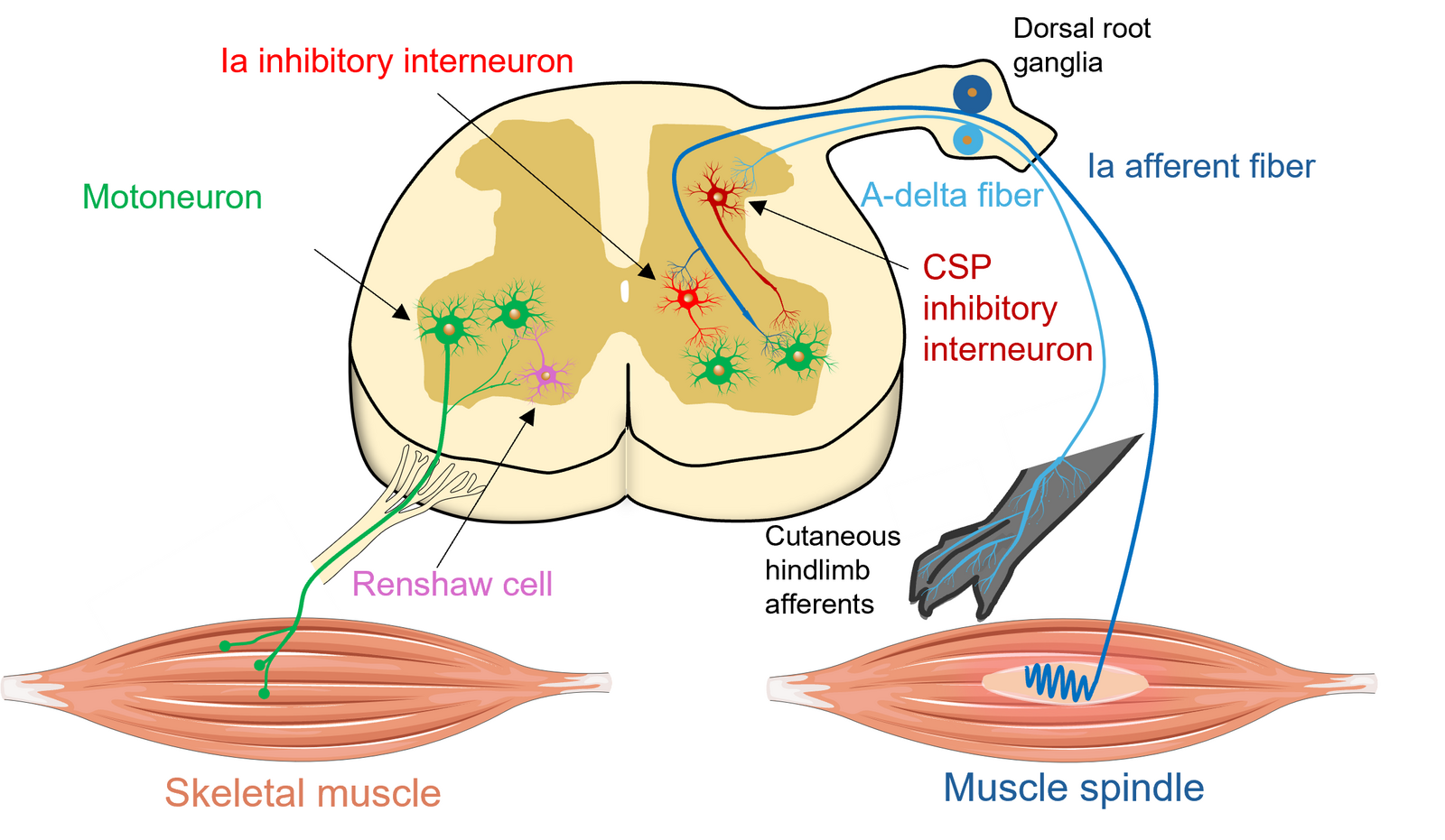
Spinal microcircuits in neuromuscular disease
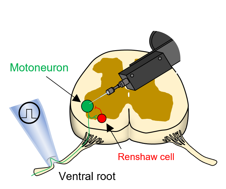
We investigate the cellular properties and synaptic function of the spinal circuits that control movement, including motoneurons and premotor interneuron populations. We examine how intrinsic properties, neuromodulatory systems and circuit connectivity shape motor output under normal conditions, and how these processes are altered in disease, particularly ALS.
In ALS, clinical weakness emerges only after more than 50% of the motoneurons have denervated a single muscle, reflecting the remarkable capacity of the central nervous system for compensation. A major goal of the lab is to identify the homeostatic adaptations in spinal microcircuits that preserve function before clinical decline, and to define windows of neuronal resilience that may be exploited for improved diagnosis, prognosis and therapeutic intervention.
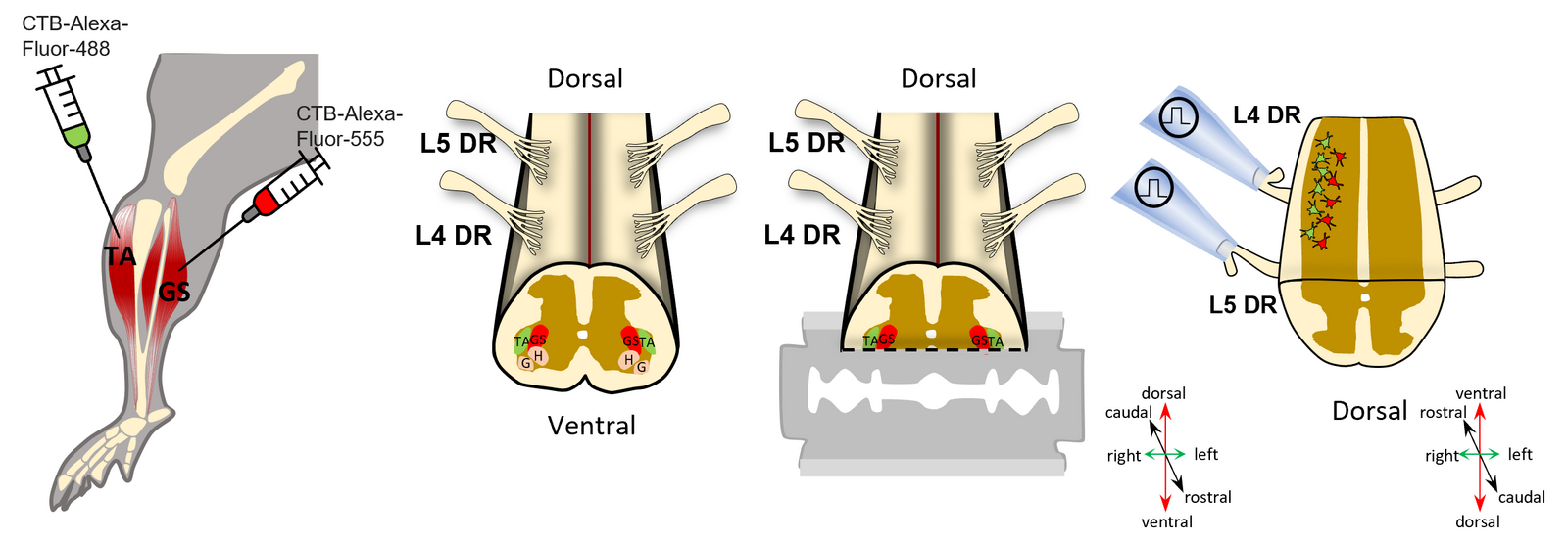
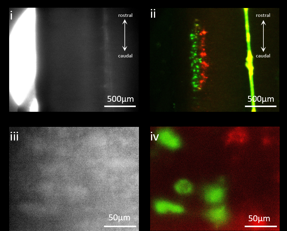
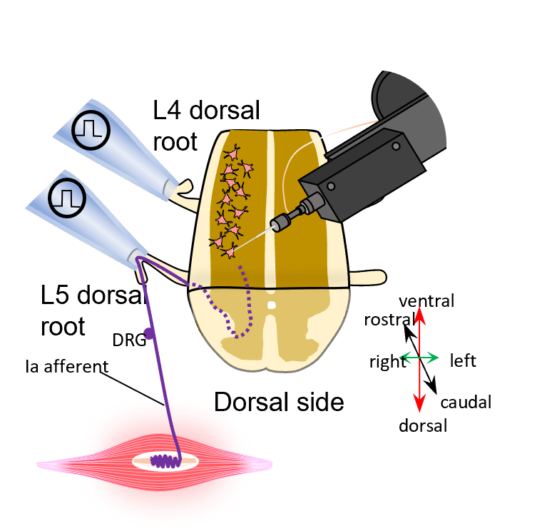
Translational neurophysiology: from mice to humans
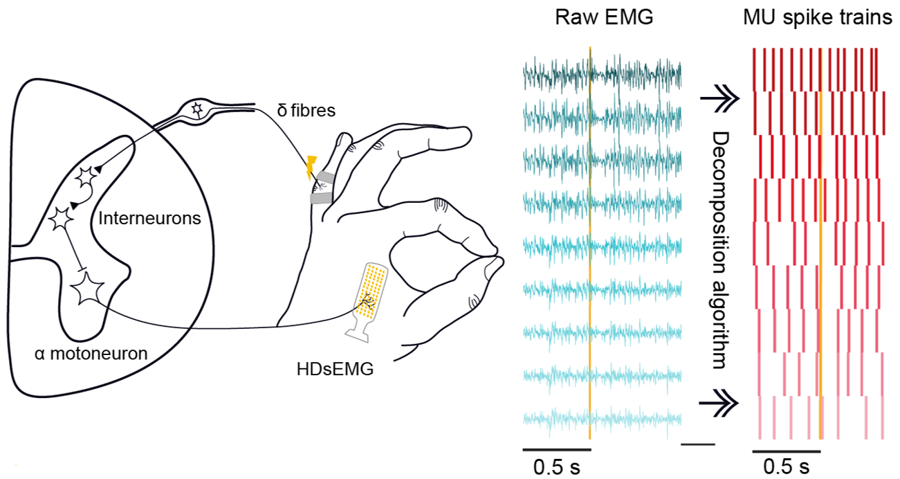
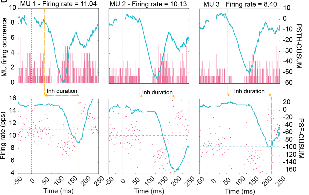
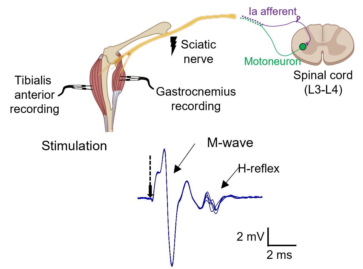
We combine in vitro, ex vivo and in vivo electrophysiology in mouse models with high-density surface electromyography (HDsEMG) in humans to study spinal microcircuits across species. The strong similarity of the neuromuscular system between species, together with directly comparable electrophysiological approaches, lets us probe mechanisms and function in parallel in mice and humans. Our goal is to identify and validate non-invasive spinal circuit biomarkers for disease staging, prognosis and longitudinal monitoring in patients.
Methods we use
In vitro electrophysiology
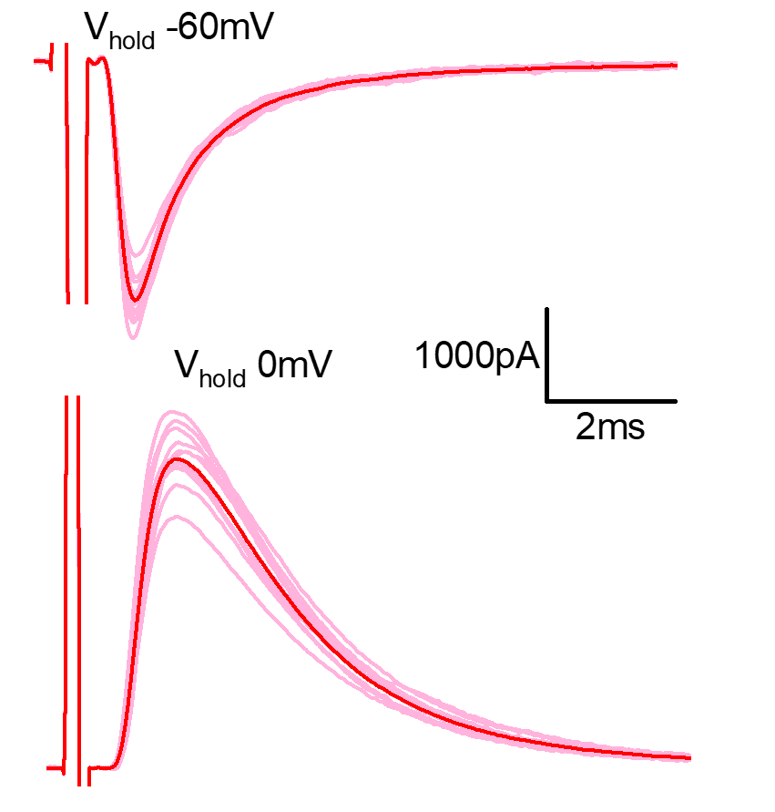
Whole-cell patch-clamp of identified motoneurons and interneurons in spinal cord slices and longitudinal preparations.
Ex vivo & in vivo recordings
Root and nerve stimulation, reflex testing and EMG in mouse models of neuromuscular disease.
HDsEMG & motor unit decoding
Non-invasive recordings of many motor units at once, used to map spinal circuit function in health and disease.
Tracing & imaging
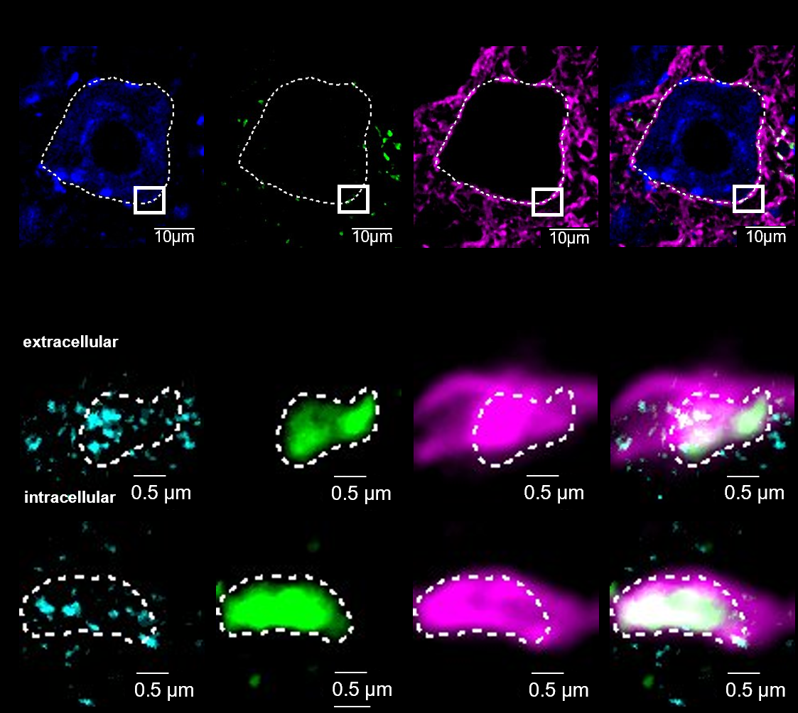
Retrograde labelling, immunohistochemistry and high-resolution microscopy of synaptic contacts.